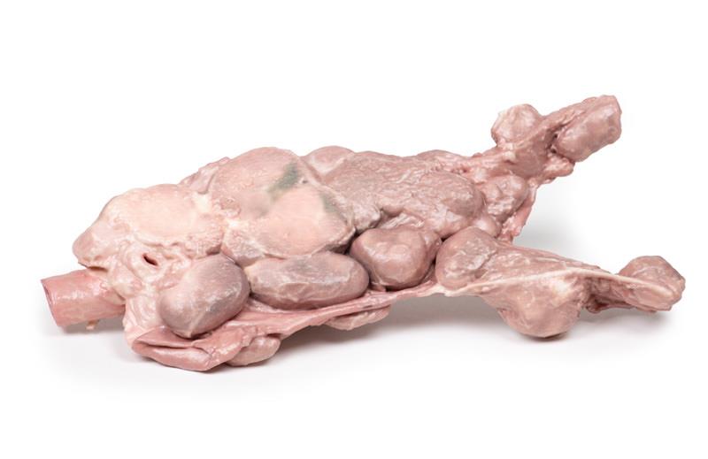
Aorta & para-aortic lymph nodes
SKU ERLMP2103
Product Description
The Aorta & para-aortic lymph nodes is a high-fidelity 3D printed anatomical model designed for healthcare professionals and educators. It replicates complex retroperitoneal structures and metastatic node patterns, helping users understand cancer recurrence and surgical strategy. This model offers a hands-on way to study challenging cases and aids in both teaching and advanced clinical planning.
Advance Medical Education with the Aorta & para-aortic lymph nodes 3D Model
This clinical-grade model brings real-life case complexity to your training, faithfully representing the abdominal aorta, common iliac arteries, and dramatically enlarged para-aortic lymph nodes. Created to mirror the high-grade adenocarcinoma metastases found in recurrent ovarian cancer, it supports accurate visualization during education or surgical simulation. With direct anatomical references, the Aorta & para-aortic lymph nodes model enables you to master both typified disease spread and the critical nuances of surgical staging, dissection, and debulking procedures. Durable 3D printing ensures consistent detail and repeated use for hands-on teaching and explant demonstration.
Features and Benefits
- Realistic 3D printed replica of abdominal aorta and iliac arteries
- Accurate representation of enlarged para-aortic lymph nodes
- Based on clinical pathology from advanced-stage ovarian cancer recurrence
- Clinical-grade detail for medical educators and surgical trainees
- Durable, reusable anatomy model for repeated handling
- Supports surgical planning, formal staging, and cancer education
- Compatible with ultrasound and PET/CT correlation studies
- Suitable for demonstration, explant teaching, and simulation
Indications for Use
- Medical teaching on lymph node metastases and disease recurrence
- Surgical simulation and planning for gynecological oncology
- Training in lymphadenectomy, dissection, and debulking techniques
- Pathology education on ovarian cancer progression
- Demonstration of imaging-pathology correlation
Size Guide
- One size – accurately scaled adult female anatomy
- Model dimensions and vascular/lymph node proportion mirror the clinical case example
You may also like
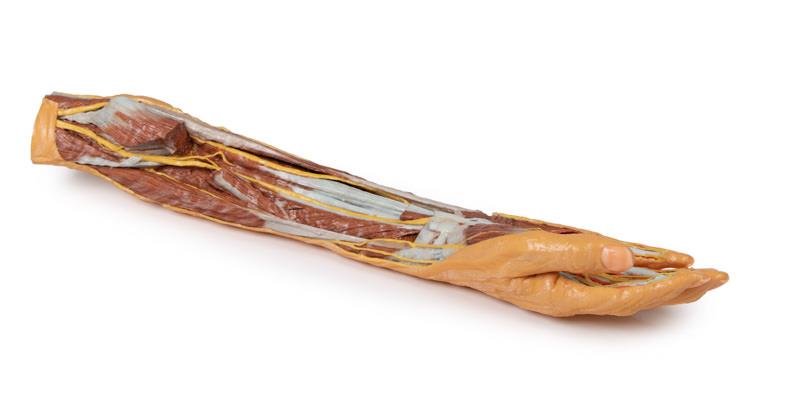
Forearm and hand - superficial and deep dissection
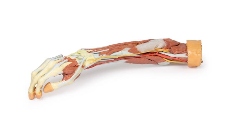
Deep upper limb and hand

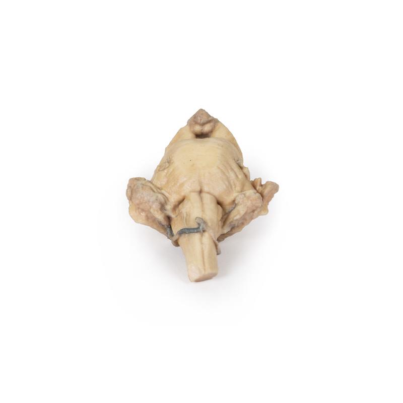
Brain stem, deep cerebral and diencephalic structures