Hydronephrosis and Hydroureter Caused by Obstruction by a Renal Calculus
SKU ERLMP2094
Product Description
The Hydronephrosis and Hydroureter Caused by Obstruction by a Renal Calculus is a detailed 3D printed anatomical model designed for medical educators, students, and healthcare professionals. It demonstrates the clinical and pathological effects of a renal calculus obstructing the urinary tract, making complex concepts easy to understand. This model offers an invaluable visual and tactile teaching aid that highlights real-life anatomical pathology.
Clear Visualization of Obstructive Uropathy Pathology
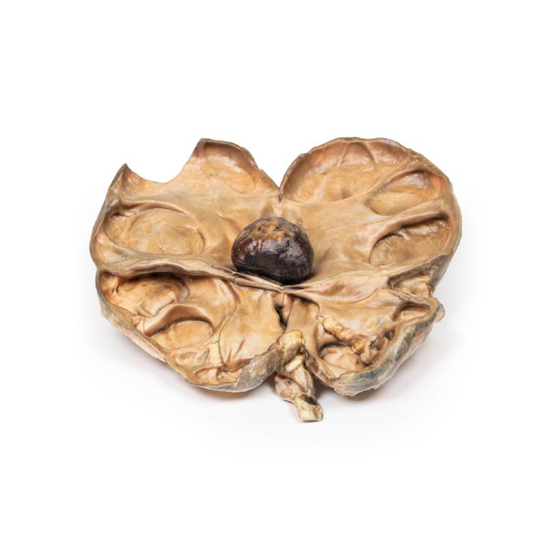
Elevate your teaching and deepen understanding with the Hydronephrosis and Hydroureter Caused by Obstruction by a Renal Calculus 3D printed model. This model features a bisected right kidney showing extensive pelvi-calyceal dilatation, renal cortical atrophy, and a prominent brown calculus lodged at the ureteropelvic junction. Use this clinical-grade model to explain disease progression, complications, and the anatomical impact of chronic obstruction in urolithiasis with accuracy and authority.
Features and Benefits
- True-to-life 3D printed representation of a right kidney obstructed by a renal calculus
- Demonstrates gross dilatation of the pelvi-calyceal system and significant loss of renal tissue in the cortex
- Highlights the presence of a large brown calculus at the ureteropelvic junction
- Shows bisected kidney anatomy for internal visualization
- Designed for clinical-grade quality, perfect for teaching and demonstration
- Durable material ensures repeated handling in classrooms and labs
- Helps visualize the effects of chronic obstruction, renal tissue atrophy, and stone formation
Indications for Use
- Medical and anatomy education in universities and teaching hospitals
- Demonstrating complications and pathology of urinary tract obstruction
- Case-based learning for nephrology, urology, and pathology studies
- Practical demonstrations for medical student and clinician training
Size Guide
- Model represents a life-size right kidney with attached ureter
- Bisected anatomy for internal detail visualization
- Dimensions follow standard adult kidney anatomy (approx. 11-12 cm length)
You may also like
Forearm and hand - superficial and deep dissection
Deep upper limb and hand

Brain stem, deep cerebral and diencephalic structures