Metastatic melanoma
SKU ERLMP2018
Product Description
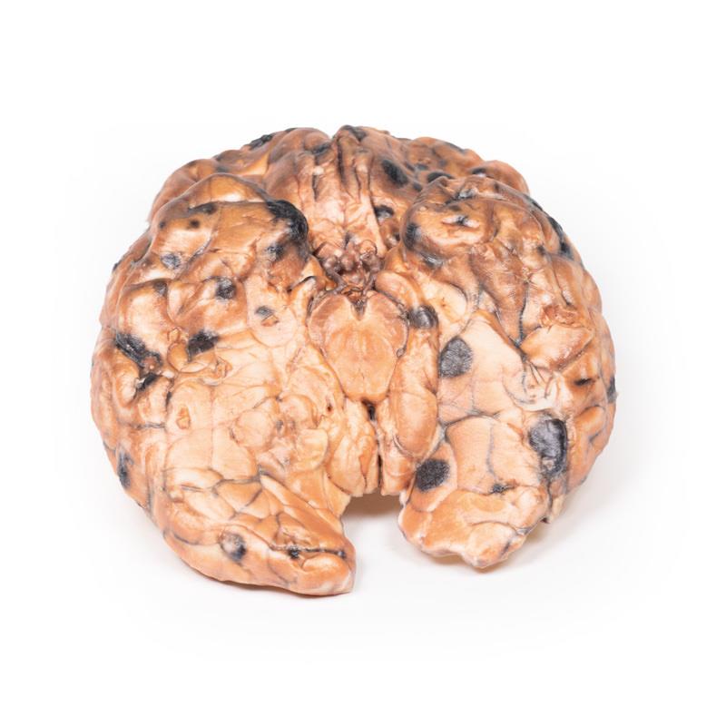
The Metastatic melanoma is a highly detailed 3D printed anatomical model designed for medical students, educators, and healthcare professionals. It provides an authentic visualisation of widespread intracerebral melanoma metastases. Choose this model to enhance medical training and understanding of metastatic disease progression, offering a real-world reference that sets it apart from generic anatomy models.
Advanced Anatomical Accuracy for Superior Education
Elevate your teaching or study sessions with the Metastatic melanoma model. Clinical-grade precision and realistic detail bring the severe implications of metastatic melanoma to life. The model replicates key pathological features, including elevated melanotic nodules and non-encapsulated tumour deposits confined to grey matter. It empowers educators, students, and clinicians to investigate neural invasion, necrosis, and haemorrhage exactly as seen in real patients. Use this model to bridge classroom theory and clinical reality, supporting critical learning for diagnosis and treatment planning.
Features and Benefits
- 3D printed replica with clinical-grade realism
- Displays multiple dark nodules up to 1.5 cm in diameter
- Highlights metastatic deposits restricted to grey matter
- Demonstrates cortical invasion and non-encapsulated tumours
- Shows areas of necrosis and haemorrhage
- Ideal for pathology, neurology, and oncology education
- Durable material suitable for repeated classroom use
- Supports neuroimaging and histopathology interpretation training
Indications for Use
- Medical and pathology education
- Patient and student demonstration of metastatic melanoma features
- Training for brain metastasis diagnosis and treatment planning
- Anatomical reference for clinicians and surgeons
Size Guide
- True-to-life scale with surface nodules up to 1.5 cm in diameter
- Represents a typical human brain specimen with metastatic melanoma involvement
You may also like
Forearm and hand - superficial and deep dissection
Deep upper limb and hand

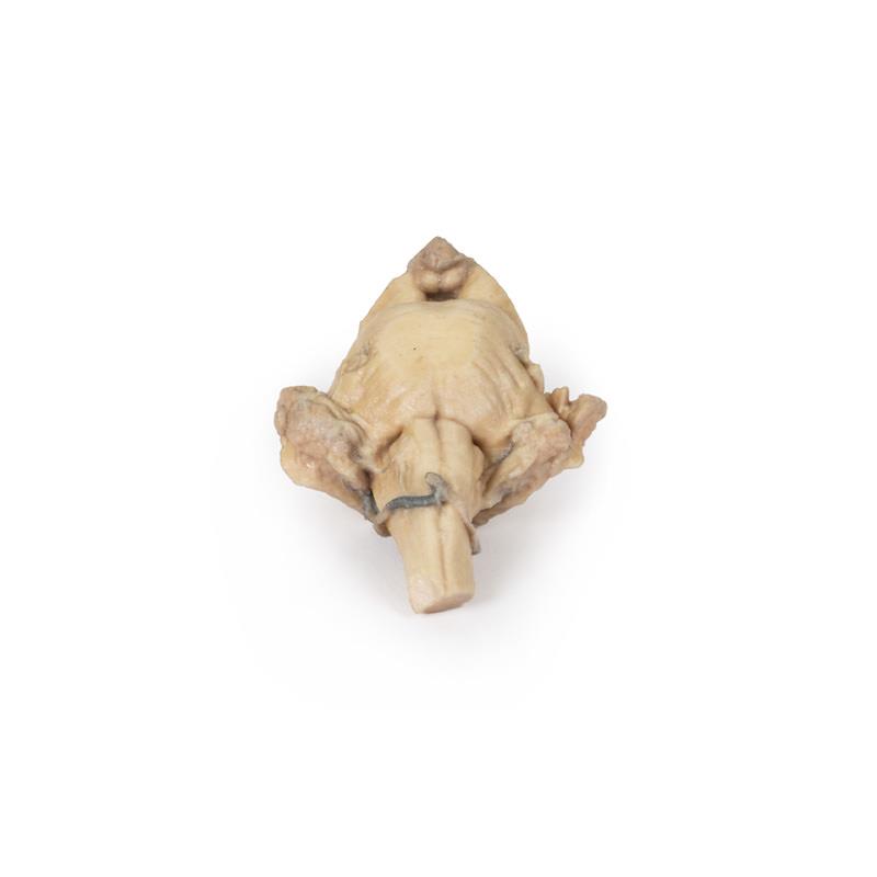
Brain stem, deep cerebral and diencephalic structures