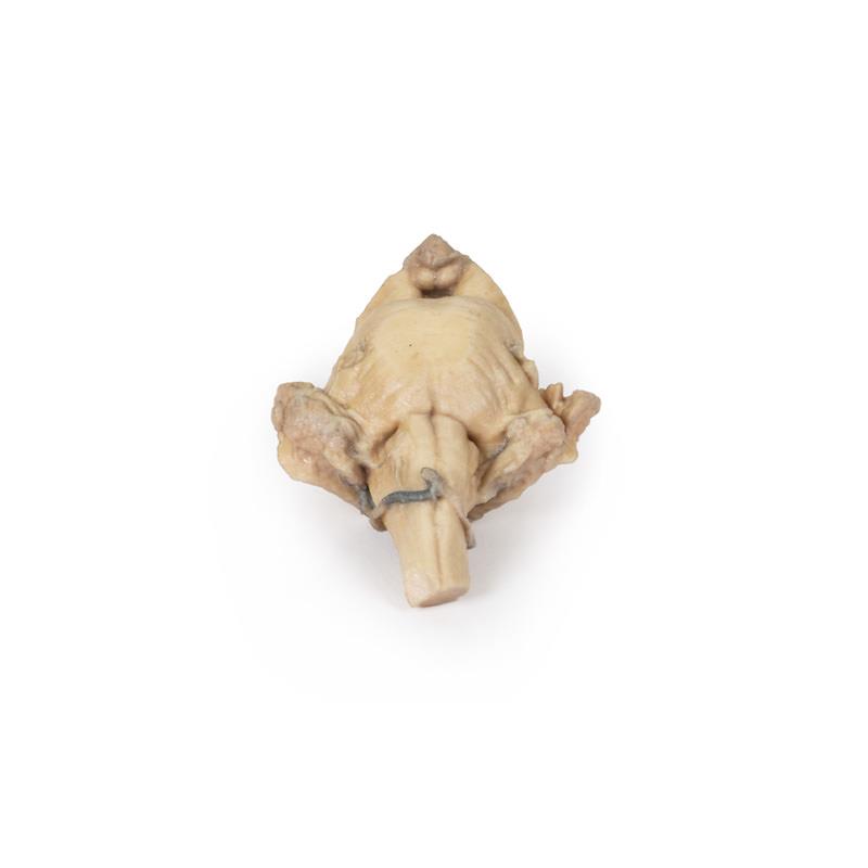

Papillary Transitional Cell Carcinoma of the Renal Pelvis
SKU ERLMP2099
Product Description
The Papillary Transitional Cell Carcinoma of the Renal Pelvis is a clinical-grade 3D printed anatomical model designed for medical educators and students. It accurately demonstrates the pathology of a papillary transitional cell carcinoma in the renal pelvis, making detailed anatomical and pathological education more effective. Select this model for true-to-life educational clarity and robust construction ideal for hands-on teaching.
Enhance Medical Training with True-to-Life Tumor Pathology
This 3D printed model vividly illustrates a post-nephrectomy kidney affected by Papillary Transitional Cell Carcinoma of the Renal Pelvis. The model shows the preserved fetal lobulation of the kidney and features a remarkably detailed 35mm friable papillary tumor within a dilated renal pelvis. Clinicians and students see actual tumor obstruction and can explore the structural effects that mirror real pathological conditions. Clinical-grade materials ensure a long-lasting model for repeated educational use, giving educators confidence when demonstrating renal tumor pathology in urology, oncology, and general anatomy courses.
Features and Benefits
- Life-sized 3D printed model with persistent foetal kidney lobulation
- Clearly displays a 35mm friable papillary tumor projecting into the renal pelvis
- Shows dilated renal pelvis from tumor obstruction
- Constructed from durable clinical-grade material for repeated handling
- Educational standard model supporting detailed pathology lessons
- Ideal for health professionals, educators, and medical students
- Fast Australian dispatch with tracked nationwide shipping
Indications for Use
- Medical anatomy and pathology teaching
- Clinical demonstration of renal pelvis tumor features
- Training in urological cancer identification
- Patient education on renal tumors and nephrectomy outcomes
Size Guide
- Life-size 3D anatomical model
- Tumor feature - 35mm diameter papillary lesion
- Represents an adult kidney post-nephrectomy
You may also like
Forearm and hand - superficial and deep dissection
Deep upper limb and hand

Brain stem, deep cerebral and diencephalic structures