3D printing has been a buzzword in healthcare for over a decade. But in anatomy education, it has quietly moved past the hype and into real, daily use. Universities and training facilities across Australia are now using 3D printed models derived from real patient CT and MRI scans to teach anatomy in ways that were not possible five years ago.
How 3D Printed Anatomy Models Are Made
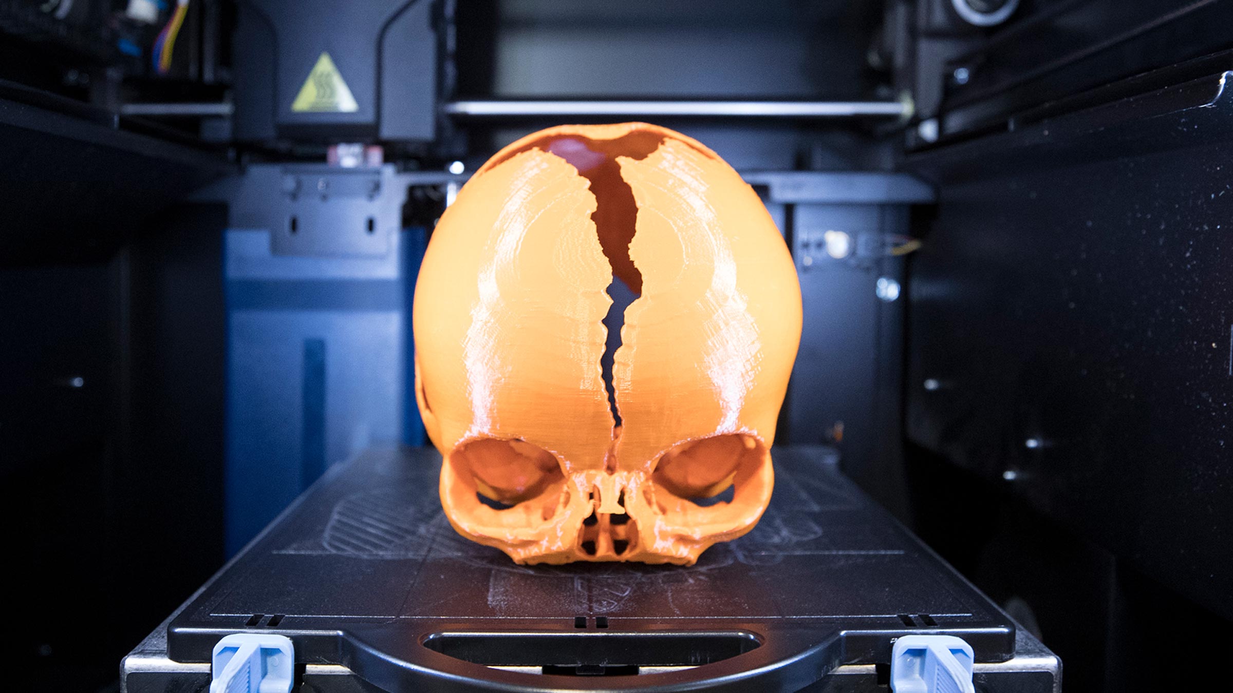
Unlike traditional anatomical models that are hand-sculpted or injection-moulded from generic references, modern 3D printed anatomy specimens start with real patient imaging data. A CT or MRI scan is segmented — meaning the structures of interest (bones, vessels, organs, pathology) are digitally isolated — and then printed layer by layer using medical-grade resins or polymers.
The result is a physical model that faithfully reproduces the exact anatomy of a real patient, including individual variations and pathological findings that generic models cannot replicate.
What Makes Them Different from Traditional Models
Patient-specific accuracy. Every 3D printed specimen reflects real human anatomy with all its natural variation. No two hearts are identical in real life, and no two 3D printed hearts need to be identical either. This teaches students to recognise and expect anatomical variation — a skill that is critical in clinical practice.
Pathology representation. 3D printed models can reproduce specific pathological findings — an abdominal aortic aneurysm, a tumour, a congenital heart defect — in a way that is impossible with standard teaching models. Students can hold, rotate, and examine a Berry aneurysm or a polycystic kidney in their hands.
Ethical sustainability. As access to cadaveric specimens becomes more restricted (due to cost, availability, health regulations, and cultural considerations), 3D printed models offer an ethical, scalable alternative. They require no preservatives, no specialised storage, and no biological hazard management.
Durability and portability. Unlike wet specimens, 3D printed models do not degrade. They can be passed around a classroom, transported between campuses, or stored on a shelf indefinitely without deterioration.
Where They Are Being Used
Australian medical schools, nursing programs, and allied health courses are integrating 3D printed models across several areas.
- Anatomy teaching — replacing or supplementing cadaveric dissection for gross anatomy courses
- Surgical planning — surgeons use patient-specific models to rehearse complex procedures before entering the operating theatre
- Patient communication — clinicians use printed models to explain diagnoses and surgical plans to patients in terms they can understand
- Assessment — anatomy practical exams (OSCEs and spotter exams) increasingly incorporate 3D printed specimens alongside traditional prosections
Limitations to Be Aware Of
3D printed models are not a complete replacement for cadaveric study. They do not replicate soft tissue texture, tissue planes, or the experience of dissection. For surgical trainees who need to understand how a scalpel moves through tissue layers, cadaveric work remains essential.
Colour accuracy can also vary between manufacturers. The best models use multi-material, multi-colour printing to distinguish arteries, veins, nerves, and parenchyma. Lower-quality prints may use uniform colours that reduce educational value.
Our Range
We carry a comprehensive range of 3D printed anatomy and pathology specimens created from real patient scan data. The collection covers cardiovascular, neurological, abdominal, musculoskeletal, and respiratory pathologies — over 100 individual specimens.
Explore our 3D printed models catalogue or contact us for recommendations based on your curriculum.