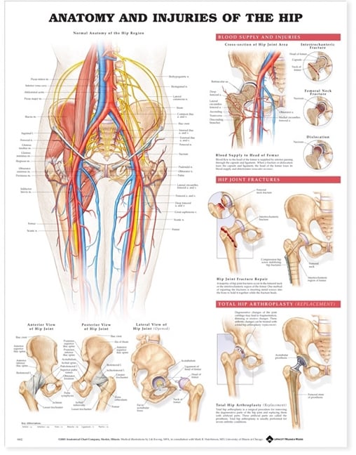
Anatomical charts and posters are a practical, low-cost teaching resource that works alongside three-dimensional models and digital tools. They give students a consistent visual reference for structures, relationships, and terminology — and they work in almost any teaching environment, from a lecture theatre to a small clinical skills room.
What Do Anatomical Charts Actually Offer in a Teaching Setting?
A well-designed anatomical chart presents a large amount of structural information in a single, readable format. Students can scan a full-body muscular chart, for example, and immediately see how muscles relate to one another across regions — something that is harder to convey with a single model or a series of slides.
Charts are also persistent. Unlike a projected slide, a laminated poster stays on the wall throughout a semester. Students walking past it between classes absorb information passively, which reinforces what they have covered in formal sessions.
For educators, charts reduce preparation time. A high-quality laminated chart of the cardiovascular system, the nervous system, or the musculoskeletal regions can anchor a lesson without requiring additional props or setup.
When Charts Work Best — and When They Fall Short
Charts are most effective for introducing terminology, reinforcing spatial relationships, and supporting self-directed revision. They are particularly useful in the early stages of a unit, when students are building a mental map of a system before they encounter models or clinical scenarios.
They are less suited to teaching three-dimensional relationships, palpation landmarks, or the tactile qualities of tissue. A chart of the shoulder joint, for instance, shows the structures clearly — but a student learning to assess shoulder pathology still needs a physical model or a clinical encounter to understand depth and movement.
The practical rule is straightforward — use charts to establish the vocabulary and the overview, then use models and simulation to build applied understanding.
Choosing the Right Charts for Your Programme
Not all anatomical charts are produced to the same standard. When selecting charts for a health-science programme, these are the key factors to weigh up —
- Accuracy and currency — the chart should reflect current anatomical terminology and be produced or reviewed by qualified anatomists.
- Lamination quality — soft laminate charts are flexible and less prone to cracking at fold lines; hard laminate is more rigid but more durable in high-traffic areas.
- Size and legibility — a chart that cannot be read from the back of a room is not useful in a lecture setting. Standard sizes range from A2 to large-format poster sizes.
- Specificity — general full-body charts suit introductory units; region-specific charts (e.g. the lumbar spine, the brachial plexus, the foot and ankle) are more useful in clinical or specialist programmes.
- Series consistency — if you are equipping a room with multiple charts, choosing from a single publisher series ensures consistent labelling conventions and visual style.
Charts and Posters in Specific Health-Science Disciplines
Different programmes use charts in different ways. Nursing and paramedicine programmes often rely on systems-based charts — cardiovascular, respiratory, neurological — to support pathophysiology teaching. A chart showing the conduction system of the heart, for example, is a useful reference when students are learning to interpret ECG rhythms.
Physiotherapy and chiropractic programmes tend to use musculoskeletal and regional charts more heavily. Charts showing muscle origins and insertions, dermatomes, or the structures of the lumbar spine are standard resources in these settings. They complement the spine and vertebrae models that students use for hands-on learning.
Medical and health-science degree programmes at Australian universities often use charts in anatomy laboratories alongside cadaveric or plastinated material. In this context, charts serve as a labelling guide and a quick reference rather than a primary teaching tool.
Veterinary programmes use species-specific charts — equine, canine, feline — to teach comparative anatomy. These are less widely available than human anatomy charts, so sourcing them from a specialist supplier is important.
Integrating Charts with Models and Simulators
The most effective anatomy teaching environments use charts, models, and simulators together rather than treating them as alternatives. A typical sequence runs as follows — students review a regional chart before a practical session, work with a physical anatomy model during the session, and then return to the chart for revision and self-testing afterwards.
This approach works because each resource type addresses a different learning need. Charts build declarative knowledge — names, locations, relationships. Models build spatial and tactile understanding. Simulators build procedural skill. Combining them reduces the cognitive load on any single resource and gives students multiple pathways to the same information.
For programmes with limited budgets, charts are often the most cost-effective starting point. A set of high-quality laminated charts covering the major body systems can be purchased for a fraction of the cost of a full model set, and they require no maintenance or storage beyond wall space.
Procurement Considerations for Australian Institutions
When purchasing charts for an Australian health-science institution, a few practical points are worth noting. Most institutions purchase charts through a single supplier to simplify invoicing and ensure consistent quality across departments. Bulk orders often attract better pricing, so it is worth consolidating requests across units or faculties before placing an order.
Laminated charts have a long service life if handled carefully. Soft-laminate charts can be rolled for storage, which makes them easier to manage in departments that rotate resources between rooms. Hard-laminate charts are better suited to permanent wall mounting.
If your programme requires charts that are not available through standard catalogues — for example, charts in a specific language, charts covering a niche anatomical region, or charts formatted to a non-standard size — it is worth contacting a specialist supplier directly to discuss options. You can also request a quote for bulk or custom orders to get accurate pricing before committing to a purchase.
For institutions that are building or refreshing a teaching space, it is also worth reviewing the full range of charts and posters available from specialist anatomy suppliers, as the range is broader than what is typically stocked by general educational suppliers.
A Practical Starting Point for New Programmes
If you are setting up an anatomy teaching space for the first time, a core set of charts covering the skeletal system, the muscular system, the cardiovascular system, the nervous system, and the respiratory system will cover the majority of introductory health-science content. From there, you can add region-specific or system-specific charts as your programme develops.
For programmes that already have a model collection, charts are a straightforward addition that requires minimal planning. Mount them at eye level in teaching spaces, ensure they are legible from a reasonable distance, and update them when new editions are released. That is the extent of the ongoing commitment — which is part of what makes them a reliable, low-maintenance teaching resource.