Eye and ear anatomy models give Australian health-science educators a hands-on way to teach sensory organ structure that diagrams and textbooks cannot replicate. This guide covers the main model types available, what to look for when selecting them, and how to match a model to your programme level and budget.
Why Eye and Ear Models Matter in Health-Science Teaching
The eye and ear are structurally complex organs with many small, closely packed components. Students who only encounter them in two-dimensional diagrams often struggle to build accurate spatial understanding of how the parts relate to one another. A physical model lets students rotate, disassemble, and reassemble the structure, which reinforces learning in a way that passive study cannot.
For programmes in optometry, audiology, nursing, paramedicine, and general health science, this kind of tactile engagement is particularly valuable. Students can trace the path of light through the lens and vitreous humour, or follow the route of sound from the outer ear through the ossicles to the cochlea, using a model they can hold and examine from every angle.
Models also support assessment preparation. When students can physically point to and name structures, they tend to retain that knowledge more reliably than when they have only labelled a diagram.
Types of Eye Models Available
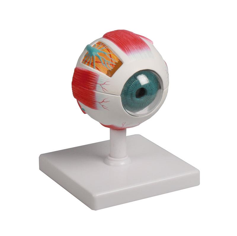
Eye models range from simple, single-piece display pieces to large, multi-part dissectible versions designed for active teaching. The most commonly used in Australian health-science programmes are enlarged dissectible models, typically four to six times life size, which allow students to remove and examine individual layers and structures.
Key features to look for in an eye model include
- Clear labelling of major structures — cornea, iris, lens, retina, optic nerve, vitreous and aqueous humour
- Removable parts that allow students to examine internal anatomy without damaging the model
- Durable construction that withstands repeated handling in a busy skills lab
- A stable base or stand so the model can be displayed between teaching sessions
- Augmented reality compatibility, where available, to extend the learning experience digitally
Some models in the Eye and Ear Models range include augmented reality features that overlay additional anatomical information when viewed through a compatible app. These can be useful for programmes that want to integrate digital tools without replacing physical models entirely.
Types of Ear Models Available
Ear models are typically divided into outer, middle, and inner ear sections, with the most detailed versions showing all three regions in a single dissectible unit. Enlarged models — often five to eight times life size — make it easier for students to identify small structures such as the malleus, incus, stapes, and cochlea that would be difficult to see at true scale.
When evaluating ear models, consider whether the model shows
- The full pathway from the pinna and external auditory canal through to the cochlea and auditory nerve
- The tympanic membrane and its relationship to the ossicles
- The semicircular canals and their role in balance
- The Eustachian tube and its connection to the nasopharynx
For programmes covering audiology or ear, nose, and throat (ENT) content, a model that shows the relationship between the middle ear and surrounding structures — including the mastoid air cells — adds teaching value beyond basic anatomy courses.
Matching a Model to Your Programme Level
The right model depends on the depth of anatomical content your programme requires. A first-year health-science unit covering sensory organ overview will have different needs from a third-year optometry or audiology programme where students need to understand pathology and clinical relevance.
For introductory courses, a straightforward enlarged model with clear colour coding and labelling is usually sufficient. Students at this level benefit from being able to identify major structures and understand basic function without being overwhelmed by fine detail.
For advanced courses, look for models with greater anatomical detail, pathology options where available, and compatibility with supplementary resources such as anatomical charts. Pairing a detailed eye or ear model with a relevant anatomical chart gives students both a three-dimensional reference and a two-dimensional overview they can use during self-directed study.
Budget and Procurement Considerations for Australian Institutions
Eye and ear models vary considerably in price depending on size, complexity, and the number of removable parts. Entry-level enlarged models suitable for introductory teaching are generally the most affordable option. Multi-part dissectible models with augmented reality features sit at the higher end of the range.
For institutions working within a fixed budget, a practical approach is to purchase one or two high-quality dissectible models for demonstration and small-group work, and supplement these with anatomical charts for individual student reference. This combination covers most teaching needs without requiring a large upfront investment in multiple physical models.
Australian institutions can request a formal quote through the quote request page, which is useful for procurement teams that need itemised pricing for budget approval or tender processes. Volume pricing may be available for larger orders.
Integrating Eye and Ear Models into Your Teaching Programme
Physical models are most effective when they are integrated into structured teaching activities rather than used as passive display pieces. Some approaches that work well in Australian health-science programmes include
- Structured identification exercises where students label structures on a diagram after handling the model
- Small-group rotation activities where each group spends time with the model before moving to a related task
- Pre-clinical assessment preparation, where students practise identifying structures under timed conditions
- Flipped classroom formats, where students review diagrams and videos before a hands-on session with the model
Models that are easy to disassemble and reassemble are particularly well suited to group activities, as students can work through the anatomy collaboratively. Robust construction is important in this context — models that are used frequently need to withstand repeated handling without parts becoming loose or breaking.
For programmes that cover both eye and ear anatomy within the same unit, purchasing models from the same product range can help ensure consistent quality and labelling conventions across your teaching resources. Browse the full anatomy models range to compare options across different organ systems.