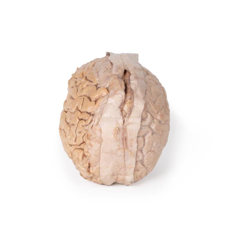
Brain anatomy models give neuroscience and health-science educators a tangible, three-dimensional reference that diagrams and textbooks cannot replicate. For Australian universities, nursing programmes, and allied health schools, a well-chosen brain model supports everything from introductory neuroanatomy lectures to clinical skills preparation. This guide covers the main model types, what to look for when selecting one, and how to match a model to your teaching context.
Why Physical Brain Models Still Matter in the Digital Age
Digital anatomy software and virtual reality tools have expanded rapidly, but physical models retain distinct advantages in a teaching environment. Students can handle a physical model without equipment, share it across a group simultaneously, and refer to it during practical assessments without a screen between them and the specimen.
Brain models are particularly useful for spatial reasoning. Understanding the relationship between the cerebral lobes, the brainstem, and the cerebellum is easier when a student can rotate a model in their hands than when they navigate a 3D interface on a flat screen. For programmes that include neurological assessment or patient communication skills, a physical model also supports role-play scenarios in a way that software cannot.
Physical models require no software licences, no charging, and no IT support. For institutions managing tight budgets or unreliable internet access, this reliability is a practical advantage.
Main Types of Brain Models Available
Brain models for educational use fall into a few broad categories, each suited to different teaching goals.
- Sectioned cerebrum models — typically two or more removable parts showing internal structures such as the corpus callosum, ventricles, and basal ganglia. Suitable for neuroanatomy units at undergraduate level.
- Full brain with brainstem — includes the cerebrum, cerebellum, and brainstem as a single or multi-part assembly. Useful for programmes covering autonomic function, cranial nerves, and reflex pathways.
- Colour-coded dissection models — lobes or functional regions are colour-differentiated to aid identification. Particularly effective for introductory courses where students are building their first mental map of brain structure.
- Pathology models — show conditions such as cerebral haemorrhage, stroke, or tumour. Relevant for nursing, paramedicine, and medical programmes where students need to recognise abnormal presentations.
- 3D printed brain models — produced from real patient scan data, these offer high anatomical accuracy and can be customised to show specific structures or pathologies. They are increasingly used in specialist postgraduate programmes.
The right type depends on the level of the course, the clinical context students are preparing for, and the budget available per unit.
Key Features to Evaluate Before Purchasing
Not all brain models are built to the same standard. When comparing options, consider the following features.
Part count and removability. A model with more removable parts allows students to explore deeper structures, but also increases the risk of parts being lost or damaged in a busy teaching environment. For large undergraduate cohorts, a two- or three-part model is often more practical than a highly detailed eight-part version.
Labelling and colour coding. Some models include numbered structures with a corresponding key; others rely on colour alone. Numbered labelling is generally more useful for assessment preparation, as students can self-test against the key without an instructor present.
Scale and material. Life-size models give students an accurate sense of proportion, which matters when they move into clinical settings. Hard resin models are more durable than soft PVC for high-use environments, though soft models can feel more realistic to the touch.
Augmented reality compatibility. Some models now include AR markers that, when viewed through a companion app, overlay additional anatomical information. This can extend the teaching value of a single model without requiring a full digital anatomy investment.
Matching Brain Models to Your Programme Level
The appropriate model complexity varies significantly between programme types.
For introductory health-science units at TAFE or first-year university level, a colour-coded two-part cerebrum model covering the four lobes, major sulci, and gyri is usually sufficient. Students at this stage are building foundational vocabulary and spatial awareness rather than detailed structural knowledge.
For undergraduate nursing and paramedicine programmes, a full brain model including the brainstem and cerebellum is more appropriate. These students need to understand cranial nerve origins, the reticular activating system, and the structures involved in common neurological emergencies such as stroke and raised intracranial pressure.
For postgraduate or specialist programmes — including neuroscience research, neurosurgery training, or advanced clinical practice — 3D printed models derived from patient imaging data offer a level of anatomical fidelity that standard manufactured models cannot match. These can be ordered to represent specific pathologies or patient populations relevant to the programme's clinical focus.
Browse the brain and head anatomy models range to compare options across these levels.
Procurement Considerations for Australian Institutions
When purchasing brain models for an Australian university or training provider, a few practical factors are worth addressing early in the procurement process.
Consider the student-to-model ratio. A single model shared across a large cohort limits hands-on time per student. For practical sessions, a ratio of no more than four to six students per model is generally recommended. Purchasing in sets rather than individually often reduces the per-unit cost.
Check warranty and replacement part availability. Models used in high-traffic teaching labs will eventually sustain damage. Suppliers who can provide replacement parts — rather than requiring a full model replacement — offer better long-term value.
For institutions that need to demonstrate value for money in procurement decisions, it is worth documenting how a model will be used across multiple units or year groups. A brain model used in neuroanatomy, clinical skills, and patient communication units across three years of a nursing programme represents a much lower cost per student contact hour than its purchase price suggests.
If your institution requires a formal quote for budget approval, you can request a quote directly through The Anatomy Store.
Caring for Brain Models in a Teaching Environment
Brain models are a long-term investment, and basic maintenance extends their useful life considerably.
Clean models with a damp cloth and mild detergent after practical sessions. Avoid alcohol-based cleaners on painted or soft PVC surfaces, as these can strip colour coding over time. Store multi-part models in their original packaging or a labelled container to prevent parts from being separated between sessions.
For models with AR markers, keep the marker surfaces clean and unscratched, as damage to the marker pattern can prevent the app from recognising the model correctly.
If a model is used for student assessment, consider designating one model as the assessment reference and keeping it in better condition than the practice models used in open lab sessions.
For a broader look at the anatomy model range available for health-science programmes, visit the anatomy models section or contact the team for advice on selecting the right models for your specific curriculum.